What Is Ga Of The Eye
What Is GA Of The Eye? Unpacking Geographic Atrophy
If you or a loved one has recently been diagnosed with a serious eye condition, you might have heard the term "GA." So, exactly what is GA of the eye? GA stands for Geographic Atrophy, and it is a major concern because it represents the advanced, severe stage of the "dry" form of Age-Related Macular Degeneration (AMD).
This diagnosis can sound scary, but understanding what GA means, how it progresses, and what steps you can take is the best way to manage the situation. This article will break down Geographic Atrophy in simple terms, discussing the symptoms, causes, and the latest treatment approaches available today.
Understanding Geographic Atrophy (GA): The Core Definition
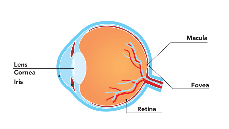
Geographic Atrophy is characterized by the clear, well-defined loss of tissue in the macula. Think of the macula as the central sweet spot of your retina, responsible for sharp, detailed central vision needed for activities like reading and driving.
When you have GA, specific cells in this area, including the retinal pigment epithelium (RPE) and photoreceptors, start to die off. This cellular death creates "geographic" regions of atrophy—areas where the tissue is completely gone—leading directly to severe, irreversible vision loss in the center of the visual field.
The Link Between GA and AMD
It is important to remember that GA is not a separate disease; rather, it is the endgame of advanced dry AMD. Age-Related Macular Degeneration generally progresses in three stages: early, intermediate, and advanced.
Dry AMD accounts for about 80-90% of all AMD cases. While most people with early dry AMD never progress to severe vision loss, approximately 10-20% of those with dry AMD will eventually develop GA. This progression is slow but steady, making regular eye exams crucial for monitoring.
How GA Affects Your Vision (Symptoms to Watch For)
The vision loss caused by GA is typically central, meaning the peripheral (side) vision often remains intact. However, the loss of central acuity can be devastating for daily life. Understanding what is GA of the eye means recognizing these symptoms:
- Difficulty reading or recognizing faces due to poor central sharpness.
- Worsening night vision and difficulty adapting to changes in light.
- The appearance of a growing, dark or blank spot (scotoma) right in the center of your view.
- A noticeable reduction in color vibrancy.
Unlike "wet" AMD, which causes rapid, sudden vision loss due to leaking blood vessels, GA usually leads to a slower, more gradual decline in vision, often taking years to cause complete central blindness.
The Mechanics Behind GA: What Is Happening Inside the Eye?
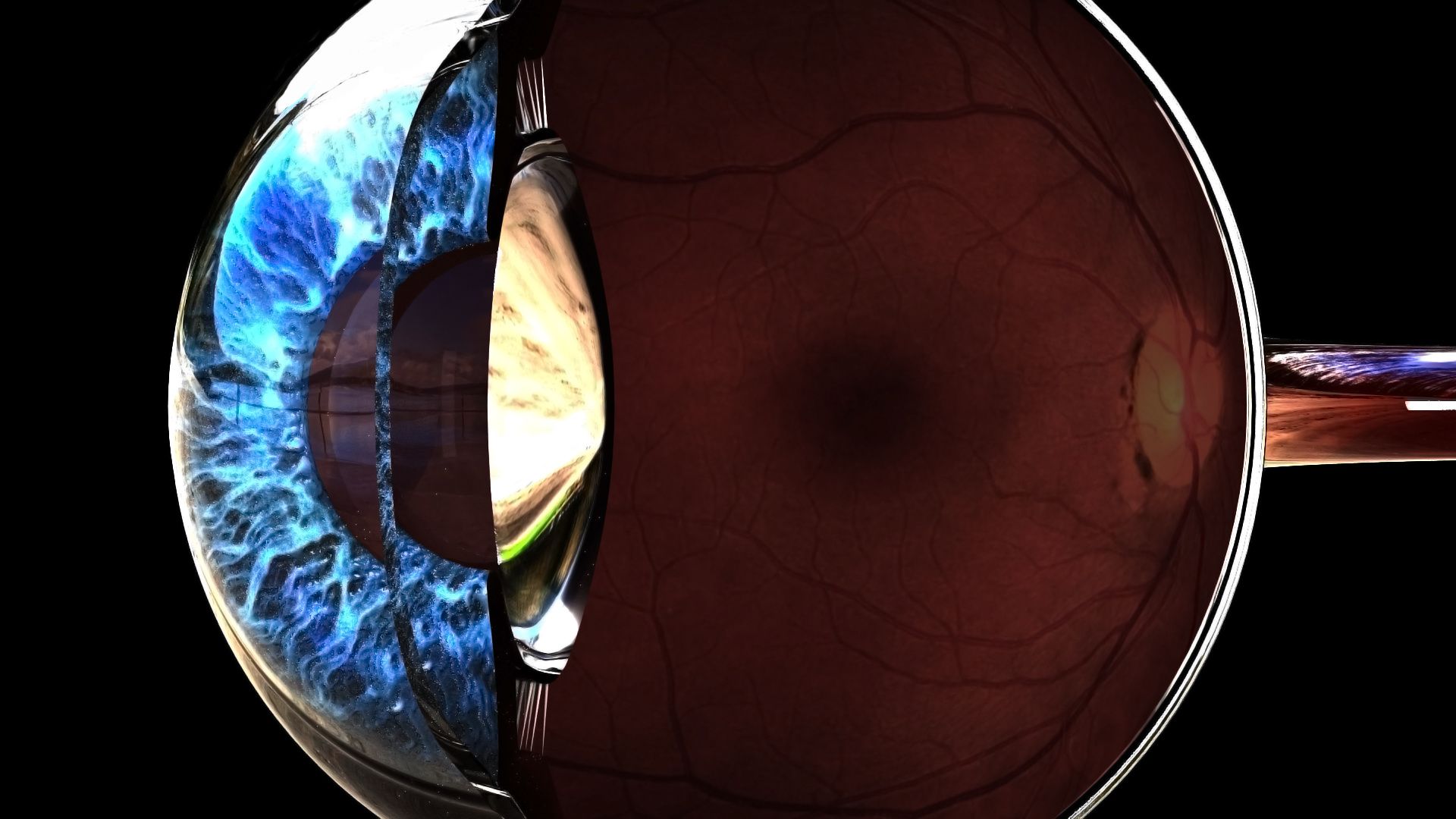
To truly answer what is GA of the eye, we need to look microscopically at the back of the eye. The process is complex, involving inflammation and chronic stress on essential retinal cells.
RPE Cells and Drusen: The Culprits
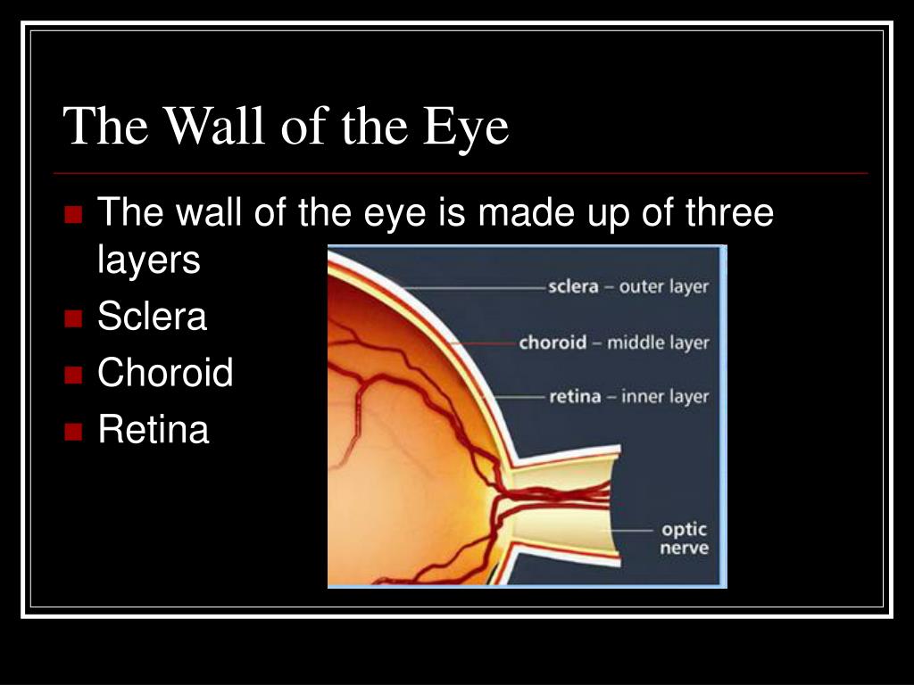
The Retinal Pigment Epithelium (RPE) layer is a crucial support system for the photoreceptors (rods and cones) which actually detect light. RPE cells recycle waste and provide nutrients. In early AMD, waste products accumulate underneath the retina, forming yellow deposits called drusen.
As drusen grow and accumulate, they stress the RPE cells. This chronic stress, coupled with inflammation and issues in the complement system (part of the body's immune defense), eventually causes RPE cells to die. Once RPE cells are gone, the photoreceptors they support also starve and die, leading to the patches of atrophy we call GA.
The Progression of Atrophy
GA typically begins in the peripheral areas of the macula, slowly moving inward toward the center (the fovea). The rate of progression varies dramatically among individuals, but on average, the atrophic lesions grow by about 1.5 to 2.5 square millimeters per year.
When the atrophy reaches the fovea, central vision loss becomes profound. Understanding this slow, relentless progression highlights why early diagnosis and potentially slowing the spread is critical.
Risk Factors: Who Is More Likely to Develop GA?
While we know what is GA of the eye, predicting exactly who will develop it remains challenging. However, several well-documented risk factors significantly increase the likelihood of progressing from dry AMD to Geographic Atrophy.
Age and Genetics
Age is, unsurprisingly, the single biggest risk factor. The condition rarely occurs before age 60, and prevalence increases dramatically with each decade thereafter. Furthermore, genetics play a huge role.
Specific genes, particularly those associated with the complement immune pathway (like CFH and C3), are strongly linked to the development of both AMD and GA. If close family members have GA, your risk is significantly higher.
Lifestyle and Environmental Factors
You have more control over these factors, making lifestyle changes vital for prevention and management.
- Smoking: Smoking is arguably the most preventable and damaging risk factor, doubling or tripling the risk of developing advanced AMD.
- Diet: A diet lacking essential nutrients, especially antioxidants like those found in dark leafy greens (lutein and zeaxanthin), can contribute to progression.
- Obesity and Cardiovascular Health: Poor overall cardiovascular health, including high blood pressure and cholesterol, is associated with a higher risk of developing advanced stages of AMD, including GA.
Diagnosis and Monitoring: Catching GA Early
Diagnosing GA requires specialized imaging tools used by retina specialists. Early detection doesn't prevent the condition, but it allows for prompt intervention to slow its progression and maximize the use of remaining vision.
Common Diagnostic Tools (OCT, Fundus Photography)
When you visit your eye doctor to check on your dry AMD, they will use several advanced technologies:
- Optical Coherence Tomography (OCT): This non-invasive scan provides cross-sectional images of the retina, allowing the doctor to measure the thickness of the retinal layers and precisely map out the areas of atrophy. This is the gold standard for defining the borders of GA.
- Fundus Autofluorescence (FAF): FAF uses the natural fluorescence of lipofuscin (a metabolic byproduct) in the RPE cells. Dying RPE cells show up as areas of reduced fluorescence, clearly outlining the geographic lesions.
- Fundus Photography: High-resolution photos are taken of the back of the eye to document the size and location of the lesions over time, helping the doctor monitor the rate of GA progression.
Treatment Options: Addressing What Is GA Of The Eye
For decades, managing Geographic Atrophy focused primarily on vision rehabilitation and slowing the initial progression from intermediate to advanced AMD using nutritional supplements.
However, the landscape has finally changed. Recent breakthroughs have given retina specialists the first-ever treatments specifically designed to slow the progression of GA itself, which answers the most hopeful aspect of what is GA of the eye treatment today.
Pharmaceutical Interventions
The latest treatments focus on inhibiting the complement system, which is a key driver of the inflammatory RPE cell death. These are intravitreal injections (injections into the eye) given regularly.
Currently, two complement inhibitors (like pegcetacoplan and avacincaptad pegol) have received approval. They don't restore lost vision, but clinical trials show they can significantly slow the rate of lesion growth by 20-30%, thus preserving usable central vision for longer periods.
Non-Pharmacological Management
In addition to these new injections, managing GA requires long-term commitment to nutritional support and visual aids:
- AREDS 2 Supplements: If you have intermediate dry AMD, taking the specific combination of high-dose vitamins and minerals outlined in the AREDS 2 study can reduce the risk of progressing to advanced GA.
- Low Vision Aids: Once significant central vision is lost, specialists can prescribe various devices, including magnifying glasses, telescopic lenses, and specialized electronic readers, to help patients maximize their remaining peripheral vision.
- Lifestyle Modifications: Maintaining a strict non-smoking status, controlling blood pressure, and adopting the Mediterranean diet remain essential support pillars.
Conclusion
Understanding what is GA of the eye means recognizing it as the advanced stage of dry AMD, characterized by the irreversible loss of central macular tissue. While the diagnosis can be daunting, remember that medical science is moving forward rapidly.
We now have specific treatments capable of slowing down the atrophy process, offering real hope for preserving vision for longer. If you have been diagnosed with GA, maintaining close communication with your retina specialist and adhering to your personalized treatment plan is the most effective strategy moving forward.
Frequently Asked Questions About Geographic Atrophy
- What is the difference between dry AMD, wet AMD, and GA?
- Dry AMD is the most common form, characterized by drusen (deposits). Wet AMD is advanced AMD involving abnormal blood vessel growth and leakage. GA (Geographic Atrophy) is the other form of advanced AMD, specifically involving the death of macular tissue.
- Can I regain the vision I lost due to GA?
- Unfortunately, vision loss caused by GA is currently irreversible because the photoreceptor cells have died. The goal of new treatments is strictly to slow down the progression of the existing lesions and prevent further loss of vision.
- Are the new treatments for GA covered by insurance?
- Since these are newer, high-cost biological treatments (complement inhibitors), coverage varies significantly based on your insurance provider and local health system guidelines. It is crucial to discuss coverage and financial assistance options with your retina specialist's office before beginning treatment.
- How often do I need monitoring if I have GA?
- Patients with GA typically require very close monitoring, often involving visits to the retina specialist every 4 to 12 weeks, especially if they are undergoing the new complement inhibitor injections. Consistent monitoring is necessary to track the progression of the atrophy and watch for potential complications.
What Is Ga Of The Eye
What Is Ga Of The Eye Wallpapers
Collection of what is ga of the eye wallpapers for your desktop and mobile devices.
Dynamic What Is Ga Of The Eye Abstract Concept
Immerse yourself in the stunning details of this beautiful what is ga of the eye wallpaper, designed for a captivating visual experience.

Exquisite What Is Ga Of The Eye Design Nature
Immerse yourself in the stunning details of this beautiful what is ga of the eye wallpaper, designed for a captivating visual experience.
Crisp What Is Ga Of The Eye Scene for Mobile
Immerse yourself in the stunning details of this beautiful what is ga of the eye wallpaper, designed for a captivating visual experience.

Gorgeous What Is Ga Of The Eye Landscape Photography
This gorgeous what is ga of the eye photo offers a breathtaking view, making it a perfect choice for your next wallpaper.

Breathtaking What Is Ga Of The Eye Design Collection
Explore this high-quality what is ga of the eye image, perfect for enhancing your desktop or mobile wallpaper.

Serene What Is Ga Of The Eye Design Digital Art
Explore this high-quality what is ga of the eye image, perfect for enhancing your desktop or mobile wallpaper.

Crisp What Is Ga Of The Eye Moment Art
A captivating what is ga of the eye scene that brings tranquility and beauty to any device.

Stunning What Is Ga Of The Eye Landscape in 4K
A captivating what is ga of the eye scene that brings tranquility and beauty to any device.

Vivid What Is Ga Of The Eye Artwork for Mobile
Experience the crisp clarity of this stunning what is ga of the eye image, available in high resolution for all your screens.

Vibrant What Is Ga Of The Eye Design Illustration
Discover an amazing what is ga of the eye background image, ideal for personalizing your devices with vibrant colors and intricate designs.

Artistic What Is Ga Of The Eye Design Photography
Find inspiration with this unique what is ga of the eye illustration, crafted to provide a fresh look for your background.

Gorgeous What Is Ga Of The Eye Abstract for Desktop
This gorgeous what is ga of the eye photo offers a breathtaking view, making it a perfect choice for your next wallpaper.

Exquisite What Is Ga Of The Eye Scene Illustration
A captivating what is ga of the eye scene that brings tranquility and beauty to any device.
Mesmerizing What Is Ga Of The Eye Image Photography
Explore this high-quality what is ga of the eye image, perfect for enhancing your desktop or mobile wallpaper.

Breathtaking What Is Ga Of The Eye Landscape Nature
Explore this high-quality what is ga of the eye image, perfect for enhancing your desktop or mobile wallpaper.

Mesmerizing What Is Ga Of The Eye Wallpaper Concept
Discover an amazing what is ga of the eye background image, ideal for personalizing your devices with vibrant colors and intricate designs.

Amazing What Is Ga Of The Eye Background for Desktop
Transform your screen with this vivid what is ga of the eye artwork, a true masterpiece of digital design.

Beautiful What Is Ga Of The Eye Background for Desktop
Experience the crisp clarity of this stunning what is ga of the eye image, available in high resolution for all your screens.

Artistic What Is Ga Of The Eye Image Nature
This gorgeous what is ga of the eye photo offers a breathtaking view, making it a perfect choice for your next wallpaper.

Gorgeous What Is Ga Of The Eye Photo in HD
This gorgeous what is ga of the eye photo offers a breathtaking view, making it a perfect choice for your next wallpaper.
Download these what is ga of the eye wallpapers for free and use them on your desktop or mobile devices.
0 Response to "What Is Ga Of The Eye"
Post a Comment